Awake Craniotomy: How Brain Surgery Helps Protect Speech, Movement, and Brain Function
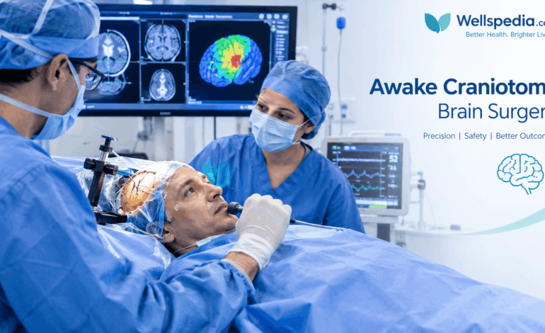
Brain surgery can be challenging when a tumor or seizure-producing area lies close to the parts of the brain responsible for speech, movement, sensation, or vision. In these situations, surgeons may recommend an advanced procedure called an awake craniotomy.
An awake craniotomy allows neurosurgeons to operate on selected brain conditions while the patient is awake for part of the procedure. During this time, the surgical team can ask the patient to speak, move a hand, name pictures, or complete other simple tasks. These responses help identify important functional areas of the brain and guide surgical decisions.
The goal is to treat the underlying condition as effectively as possible while reducing the risk of neurological damage. Although awake brain surgery offers important advantages for carefully selected patients, it is not suitable for everyone and requires an experienced, coordinated medical team.
Key Takeaways
- Awake craniotomy is a specialized form of brain surgery in which the patient is conscious during the brain-mapping portion of the operation.
- It may be considered for certain brain tumors and epilepsy-related conditions located near important functional areas.
- Electrical stimulation and real-time tasks can help surgeons identify regions involved in speech, movement, and sensation.
- Anesthesia is carefully adjusted to support comfort, safety, and cooperation during surgery.
- Potential benefits include more precise surgical planning and a better chance of preserving important neurological functions.
- Risks include seizures, bleeding, infection, swelling, and temporary or permanent neurological changes.
What Is an Awake Craniotomy?
An awake craniotomy is a neurosurgical procedure in which part of the skull is temporarily opened to provide access to the brain, while the patient is awake during a planned phase of the operation.
It is commonly considered when a brain tumor or an area responsible for epileptic seizures is located close to regions that control essential functions.
The brain contains specialized networks responsible for activities such as speaking, understanding language, moving the limbs, processing sensations, and interpreting visual information. These networks can vary from one person to another. A tumor or neurological condition may also change how nearby brain regions function.
Brain imaging helps surgeons plan an operation, but imaging alone cannot always establish exactly how an individual brain region contributes to a specific task. Awake brain mapping allows the surgical team to assess function directly during the procedure.
Why Is the Patient Awake During Surgery?
The awake phase gives the surgical team an opportunity to observe how the brain responds while the patient performs simple activities.
For example, a patient might be asked to name an everyday object displayed on a screen. If stimulation of a particular brain region repeatedly interferes with naming, that region may be important for language.
Similarly, the team may ask the patient to move a finger or foot while testing selected areas. This helps identify regions that contribute to movement.
The information guides the surgeon in deciding where to operate and which areas should be preserved whenever possible.
Important: The patient is not necessarily awake throughout the entire operation. Anesthesia and sedation are tailored to the procedure, the patient’s needs, and the medical team’s approach.
Why Is Awake Brain Surgery Performed?
The main purpose of awake craniotomy is to treat a brain abnormality while protecting essential neurological functions.
1. Brain Tumor Removal
Some brain tumors, including certain gliomas, develop near areas responsible for speech, movement, or other important abilities.
Removing as much tumor tissue as safely possible may be an important treatment goal. However, aggressive removal can cause neurological problems if critical functional tissue is damaged.
Awake mapping provides real-time information that can help surgeons balance tumor removal with preservation of function. It does not eliminate all risks, and the appropriate extent of removal depends on the tumor’s location, biology, and the patient’s overall condition.
2. Epilepsy Surgery
Some people with epilepsy continue to experience seizures despite appropriate medication. When investigations identify a particular brain region responsible for the seizures, surgery may be considered.
Awake mapping can help in selected cases where the suspected seizure-producing area is close to essential functional regions. The aim is to treat the seizure focus while minimizing harm to surrounding brain networks.
3. Surgery Near Language and Movement Areas
Awake craniotomy may be particularly valuable when a lesion lies close to regions involved in speaking, understanding language, or controlling movement.
The surgical team can use the patient’s responses to help make decisions during the operation rather than relying only on preoperative images.
4. Individualized Surgical Planning
The exact approach depends on the location of the abnormality, the patient’s neurological abilities, medical history, and the goals of treatment.
Not every brain tumor or epilepsy operation requires an awake procedure. Conventional surgery under general anesthesia may be more appropriate in many cases.
Who May Be Suitable for an Awake Craniotomy?
Awake craniotomy is generally considered when the expected benefit of direct functional mapping outweighs the additional demands of the procedure.
Factors that may support its use include:
- A brain lesion close to important language or motor areas.
- A need to identify functional boundaries during tumor removal.
- An epilepsy-related surgical plan that requires functional mapping.
- The ability to understand and follow instructions during the awake phase.
- A medical condition and airway plan compatible with the proposed anesthesia technique.
The team must also consider anxiety, communication difficulties, cognitive impairment, breathing problems, seizure risk, and other factors that may affect safety or cooperation.
A neurosurgeon and neuro-anesthesia team assess these issues before recommending the procedure.
How Is an Awake Craniotomy Performed?
The precise sequence varies between hospitals and patients. Many operations use an anesthesia technique that allows the patient to be asleep or deeply sedated during some stages and awake during brain mapping.
Step 1: Preoperative Assessment
Before surgery, the medical team reviews brain imaging, the planned surgical target, and the patient’s existing neurological function.
Depending on the case, assessments may include:
- Neurological examination.
- Speech and language evaluation.
- Movement and sensory testing.
- Cognitive assessment.
- MRI and other relevant imaging.
- Anesthesia assessment and airway planning.
The patient is also informed about what to expect during the awake phase and which tasks may be required.
Step 2: Positioning and Anesthesia
The patient is positioned to give the surgical team safe access to the target area. The head is secured to help prevent movement during delicate surgical work.
Local anesthetic is used to numb relevant scalp tissues. Sedation or general anesthesia may be used during selected stages, depending on the planned technique.
The anesthesia team closely monitors breathing, oxygen levels, circulation, comfort, and responsiveness throughout the operation.
Step 3: Opening the Skull
The surgeon makes an incision in the scalp and temporarily removes a section of skull to access the brain. This opening is called a craniotomy.
When the procedure requires awake mapping, the team carefully manages the transition to a sufficiently alert state before testing begins.
The brain itself does not have the same pain-sensitive structures as the scalp, although surrounding tissues can cause discomfort. Appropriate anesthesia and monitoring are therefore essential.
Step 4: Brain Mapping and Functional Testing
The surgeon may apply brief electrical stimulation to selected brain areas while a specialist asks the patient to perform specific tasks.
These tasks may include:
- Naming pictures or familiar objects.
- Repeating words or sentences.
- Reading short passages.
- Following spoken instructions.
- Moving a finger, hand, or foot.
- Reporting sensations such as tingling.
The team records any changes in performance and uses these observations to build a functional map.
Step 5: Treating the Target Area
Once relevant functional areas have been identified, the surgeon proceeds with the planned treatment.
For a tumor, this may involve removing tissue while avoiding or limiting damage to important functional networks. In epilepsy surgery, the procedure may target a seizure-producing region according to the surgical plan.
Brain mapping can guide decisions, but it cannot guarantee that all essential functions will remain unchanged.
Step 6: Closing the Surgical Site
After the necessary work is complete, the surgical team closes the skull opening and scalp incision. The patient may receive additional sedation or anesthesia if needed.
Following surgery, the healthcare team monitors neurological function, breathing, pain, and other signs of recovery.
What Tests Are Performed During Awake Brain Mapping?
The tests used during awake craniotomy depend on which brain functions are most at risk. A multidisciplinary team may include neurosurgeons, neuro-anesthesiologists, neurologists, and speech-language specialists.
Motor Mapping
Motor mapping helps identify brain areas involved in controlling movement.
The patient may be asked to move a hand, finger, arm, or foot. Electrical stimulation of selected areas can alter movement or produce a muscle response. The team records these findings to help identify regions that should be protected during surgery.
This information is particularly important when a lesion is near the motor cortex or the pathways that carry movement signals.
Sensory Mapping
Sensory mapping evaluates regions involved in processing physical sensations.
During testing, the patient may be asked to describe sensations such as tingling or changes in touch. These responses can help the team identify areas associated with sensory function.
The specific tests used depend on the location of the lesion and the functions that need protection.
Speech and Language Mapping
Language involves multiple brain networks rather than a single isolated center. These networks may be organized differently between individuals, and a person’s dominant language hemisphere cannot always be assumed from handedness alone.
During mapping, a speech-language specialist may ask the patient to:
- Name familiar objects shown in pictures.
- Repeat words or sentences.
- Read aloud.
- Describe a picture.
- Follow verbal instructions.
- Answer simple questions.
If stimulation interrupts speech or causes repeated naming errors, the finding may indicate that the tested region contributes to language function.
People who speak more than one language may need testing in multiple languages when clinically appropriate.
Visual Pathway Assessment
When surgery is near visual pathways, the team considers the risk of affecting eyesight or visual fields.
Visual function may be assessed through preoperative examinations, imaging, and specialized intraoperative methods when appropriate. Not every visual pathway can be mapped using the same awake tasks, so the approach is individualized.
Cognitive and Other Functional Tasks
Depending on the surgical location and clinical goals, selected procedures may include tasks involving attention, memory, counting, or other cognitive abilities.
These assessments are chosen carefully because the patient must remain comfortable and able to perform the tasks reliably.
Benefits of Awake Craniotomy
When appropriately selected and performed by an experienced team, awake craniotomy can offer several potential benefits.
More Direct Functional Information
Real-time testing helps the team identify areas associated with particular neurological functions. This may provide information that cannot be obtained from structural imaging alone.
Support for Safer Tumor Removal
Functional mapping may help surgeons remove more tumor tissue when appropriate while limiting injury to essential brain networks. The actual amount of tissue that can be removed safely varies between cases.
Preservation of Speech and Movement
Testing during surgery can alert the team when an operative step affects language or motor performance. This information can help guide decisions intended to reduce neurological injury.
Assistance With Selected Epilepsy Procedures
For some patients undergoing epilepsy surgery, functional mapping helps the team consider how treatment of the seizure-producing region may affect speech, movement, or other abilities.
Personalized Surgical Decisions
Awake mapping allows surgical decisions to be informed by the patient’s own functional responses. This can be especially useful when the lesion lies near areas with a high risk of causing disability if damaged.
These benefits are potential rather than guaranteed. Outcomes depend on the condition being treated, the location and extent of the lesion, the patient’s health, and the expertise of the treating team.
Challenges and Risks of Awake Craniotomy
Awake craniotomy requires careful planning because the patient must remain medically stable and sufficiently alert to participate during the mapping phase.
Challenges During the Awake Phase
Possible difficulties include:
Anxiety or fear: Being aware during part of brain surgery can be stressful. Preoperative explanation, reassurance, and support from the team may help.
Pain or discomfort: Although the brain tissue itself does not sense pain in the same way as skin, the scalp and surrounding tissues can be sensitive. Local anesthesia and other comfort measures are important.
Nausea: Some patients experience nausea or vomiting, which the anesthesia team works to prevent and manage.
Fatigue or difficulty following instructions: Tiredness, medication effects, or the length of the procedure can make tasks more difficult.
Seizures: Electrical stimulation can sometimes trigger a seizure. The surgical team is trained to recognize and manage this complication.
Breathing or circulation problems: The anesthesia team monitors the patient closely and responds to changes in breathing, oxygen levels, or blood pressure.
If the patient cannot safely or reliably participate, the team may need to modify the plan.
Possible Complications After Surgery
Awake craniotomy carries many of the same risks as other forms of brain surgery, including:
- Bleeding or a blood clot.
- Infection.
- Brain swelling.
- Seizures.
- Temporary or persistent weakness.
- Speech or language difficulties.
- Sensory changes.
- Visual field problems.
- Changes in memory or other cognitive abilities.
- Complications related to anesthesia.
The type and likelihood of complications vary depending on the underlying condition, the surgical location, and the patient’s health. A detailed discussion with the treating neurosurgeon is essential before making a decision.
Recovery After an Awake Craniotomy
Recovery differs from one patient to another. Some people remain in a specialized monitoring unit initially, while others may need intensive care depending on their condition and the complexity of the operation.
The healthcare team assesses neurological function and watches for early signs of complications.
What Happens Immediately After Surgery?
Initial care may include:
- Repeated checks of speech, strength, alertness, and sensation.
- Monitoring of breathing, blood pressure, and other vital signs.
- Pain and nausea management.
- Follow-up brain imaging when clinically indicated.
- Assessment for seizures or other complications.
The timing of discharge depends on the patient’s recovery and medical needs.
Rehabilitation and Follow-Up
Some patients benefit from physical therapy, occupational therapy, or speech and language therapy after surgery.
Follow-up appointments allow the team to review recovery, discuss pathology or other test results, and plan further treatment when necessary. Depending on the diagnosis, additional treatment may include medication, radiation therapy, chemotherapy, or continued epilepsy management.
Warning Signs After Discharge
Follow the hospital’s discharge instructions and seek urgent medical attention for new or worsening weakness, difficulty speaking, seizures, confusion, severe or worsening headache, fever with concerning symptoms, repeated vomiting, or reduced consciousness.
Sudden severe neurological symptoms require emergency assessment.
Awake Craniotomy Compared With Traditional Brain Surgery
Both awake craniotomy and conventional craniotomy involve opening the skull to access the brain. The main difference is how anesthesia and functional assessment are managed.
| Feature | Awake craniotomy | Conventional craniotomy |
|---|---|---|
| Anesthesia | Patient is awake during a planned phase | Usually performed under general anesthesia |
| Functional testing | Real-time tasks may be used during surgery | Assessment relies on the planned methods for that procedure |
| Common consideration | Lesions near selected functional brain regions | Many types of brain conditions requiring surgery |
| Patient participation | Required during the mapping phase | Not usually required during the operation |
| Suitability | Depends on the lesion, patient, and team | Depends on the condition and surgical plan |
Neither method is universally better. The appropriate approach depends on the condition being treated and the safest way to achieve the intended surgical goal.
Frequently Asked Questions
Is awake craniotomy painful?
The procedure is designed to control pain and discomfort using local anesthesia and carefully managed sedation. Some patients experience pressure, discomfort, anxiety, or fatigue during the awake phase. The team monitors the patient and provides appropriate support.
Is the patient awake for the entire brain operation?
Not necessarily. Some approaches use an asleep-awake-asleep technique, while others use different anesthesia plans. The patient is awake during the period when functional testing is needed, not automatically throughout the entire operation.
Why do surgeons ask patients to speak during brain surgery?
Speaking and naming tasks help the team identify brain regions involved in language. If stimulation of an area repeatedly disrupts speech, the surgeon can use that information when deciding how to proceed.
Is awake craniotomy safer than conventional brain surgery?
Awake craniotomy may provide valuable functional information for selected operations, but it is not risk-free. Whether it is the safest or most effective approach depends on the patient’s condition, the lesion’s location, and the team’s assessment.
Can a patient have a seizure during awake brain surgery?
Yes. Electrical stimulation can occasionally trigger a seizure during mapping. The surgical team prepares for this possibility and follows established protocols to manage it.
How long does an awake craniotomy take?
The duration varies according to the type and location of the lesion, the complexity of mapping, and the amount of treatment required. The surgical team can provide an estimate for an individual case.
Will speech or movement return to normal after surgery?
Many patients recover well, but outcomes differ. Some experience temporary neurological changes, while others may have persistent difficulties. The likelihood of recovery depends on the condition being treated, the brain regions involved, and other clinical factors.
Is awake craniotomy used for every brain tumor?
No. It is mainly considered when direct functional mapping is expected to provide important information. Many brain tumors can be treated using other surgical approaches, depending on their location and characteristics.
Conclusion
Awake craniotomy is a specialized neurosurgical technique that allows surgeons to assess selected brain functions while treating certain tumors and epilepsy-related conditions. By combining careful anesthesia, electrical stimulation, and real-time patient tasks, the surgical team can build a more detailed understanding of the brain areas that need protection.
The procedure may help balance effective treatment with preservation of speech, movement, and other important abilities. However, it requires careful patient selection, experienced specialists, and a clear plan for postoperative monitoring and rehabilitation.
Anyone considering brain surgery should discuss the available approaches, expected benefits, potential complications, and recovery plan with a qualified neurosurgical team.
For more evidence-informed health information and related neurological topics, explore Wellspedia.com.
Medical Disclaimer: This article is intended for educational purposes only and does not replace professional medical advice, diagnosis, or treatment. Surgical decisions must be made with qualified healthcare professionals who understand the individual patient’s condition.